
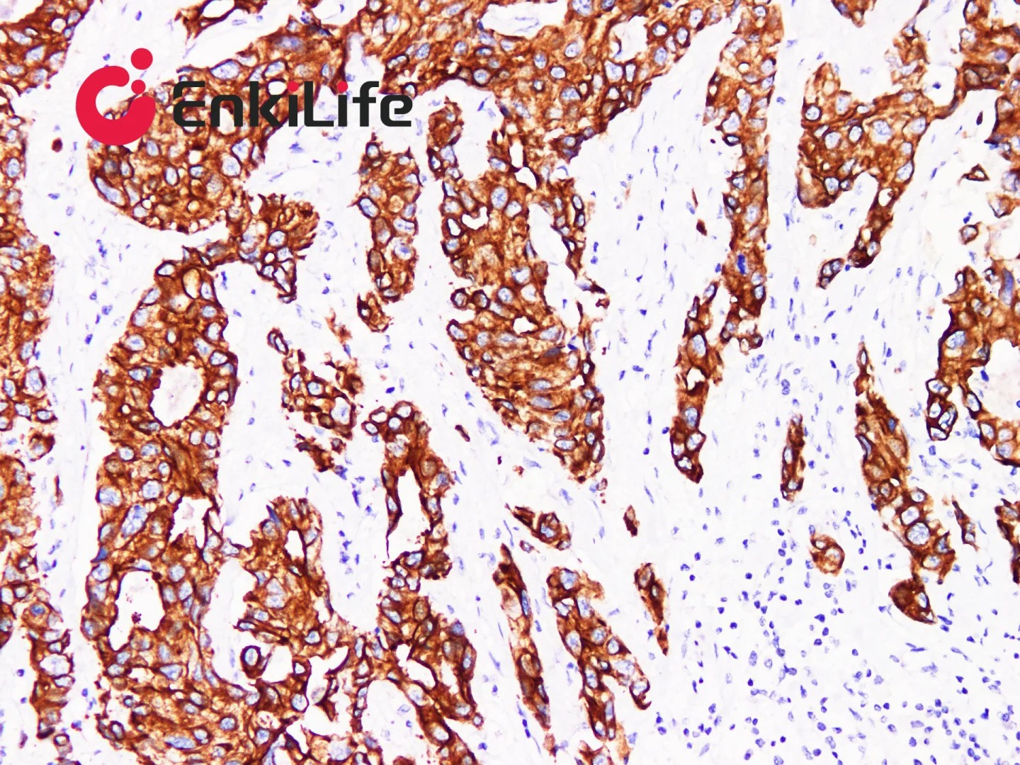
特异性识别到可视化输出:IHC 信号放大体系及检测技术详解
免疫组织化学(Immunohistochemistry, IHC)是融合免疫学、组织化学与显微成像的经典生物检测技术。核心依托抗原‑抗体特异性结合原理,结合化学显色反应,实现组织细胞内目标蛋白的原位定性、定位与半定量分析,是生命科学研究与临床病理诊断的核心技术之一。
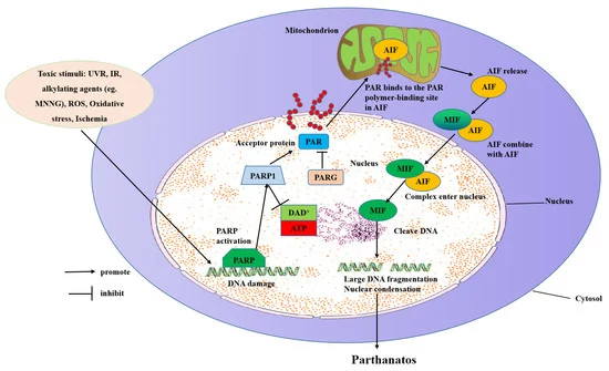
Parthanatos is a regulated cell death triggered by poly(ADP-ribose) (PAR) accumulation, driven by overactivated PARP-1. Excessive PAR binds AIF, which complexes with MIF, translocates to the nucleus, and cleaves DNA, causing cell death. It is closely linked to neurodegenerative diseases like Alzheimer’s and Parkinson’s.
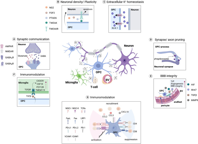
cMicroglia, as the resident immune cells in the central nervous system, exhibit distinct activation states under pathological conditions such as neurological diseases and aging, which are closely associated with neurodegeneration, inflammatory response and other processes. This paper systematically elaborates the classification of microglial activation states identified by RNA-seq technology, and summarizes the core molecular markers of microglia, including CD68, Galectin-3, Cathepsin B, HIF-1α and Axl, as well as their biological functions in phagocytosis, inflammatory regulation and neuronal injury. Meanwhile, a variety of commercial antibodies suitable for experimental detection of the above markers are collated, covering their application scenarios such as Western blot (WB), immunohistochemistry (IHC) and immunofluorescence (IF). This study provides a reference for the selection of specific detection tools and related basic research on microglial activation in neurological disease models.
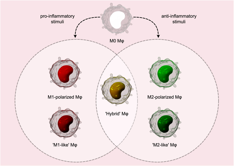
Macrophages are myeloid cells of the innate immune system that are found in all human tissues in the body and exhibit anatomical and functional diversity. These heterogenous cells are derived from monocyte precursors in the blood that infiltrate into the tissues and differentiate in the presence of cytokines and growth factors.
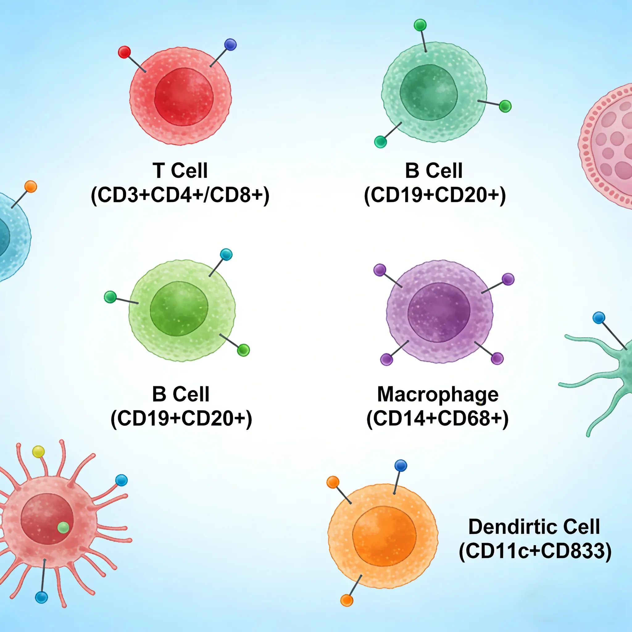
Cluster of Differentiation 3 (CD3) is a multiunit protein complex expressed on the surface of T cells that directly associates with the T cell receptor (TCR). CD3 is composed of four polypeptides: ζ, γ, ε and δ. Engagement of the TCR complex with antigens presented in Major Histocompatibility Complexes (MHC) induces tyrosine phosphorylation in the immunoreceptor tyrosine-based activation motif (ITAM) of CD3 proteins.
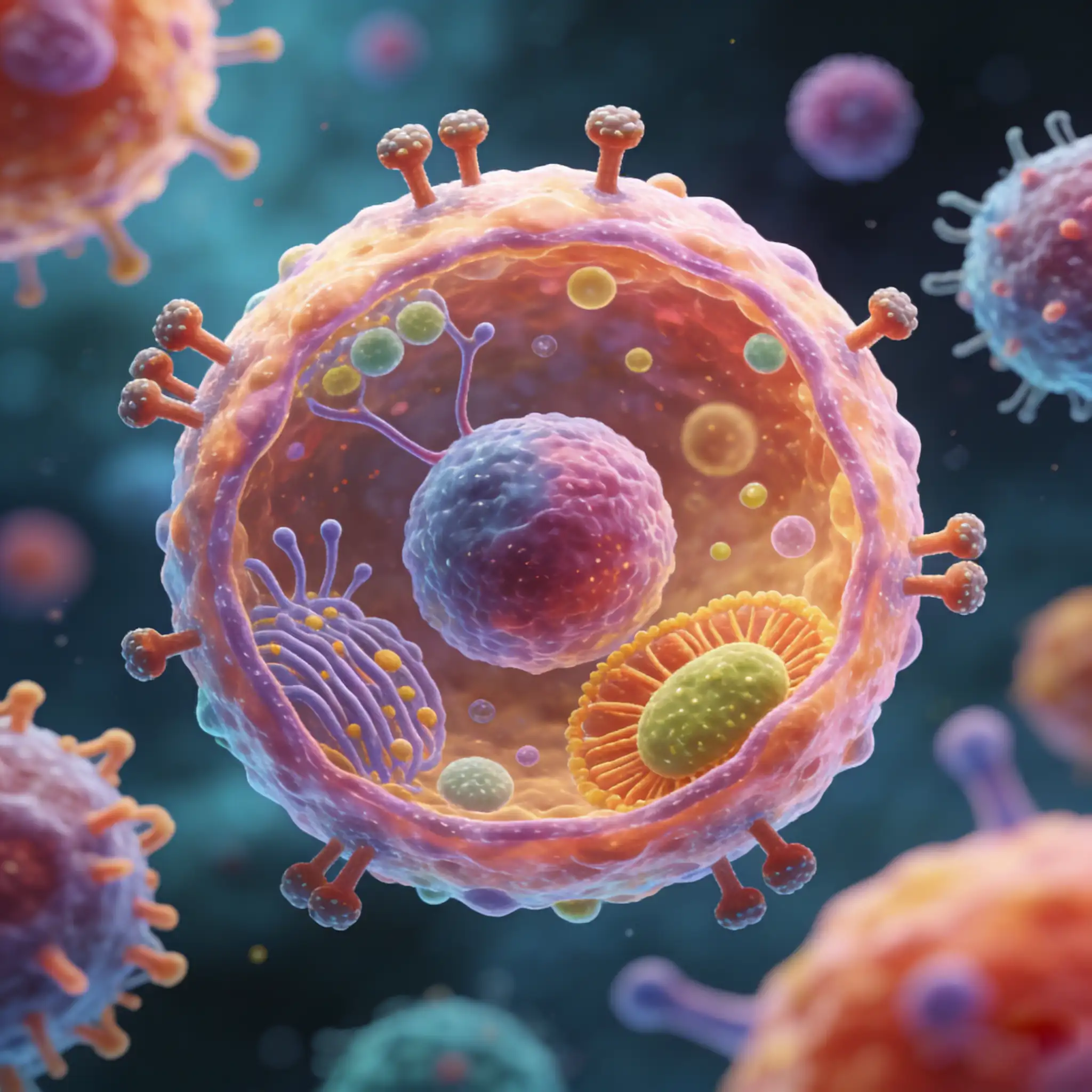
Cluster of Differentiation 3 (CD3) is a multiunit protein complex expressed on the surface of T cells that directly associates with the T cell receptor (TCR). CD3 is composed of four polypeptides: ζ, γ, ε, and δ. Engagement of the TCR complex with antigens presented in major histocompatibility complexes induces tyrosine phosphorylation in the immunoreceptor tyrosine-based activation motif (ITAM) of CD3 proteins. CD3 phosphorylation is required for downstream signaling through ZAP-70 and p85 subunit of PI-3 kinase, leading to T cell activation, proliferation, and effector functions (1).
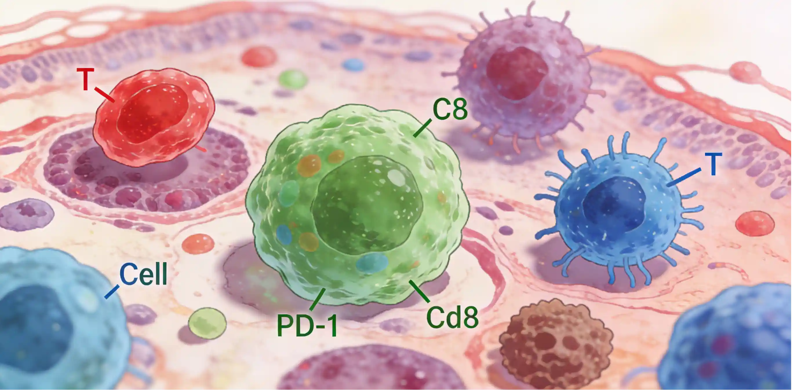
The immune system has the capability to detect and eliminate tumor cells, most notably through cytotoxic CD8+ T cell recognition of tumor antigens presented on the surface of tumor cells by MHC class I molecules. However, anti-tumor immune cells can be subject to various immunosuppressive mechanisms in the tumor microenvironment (TME).
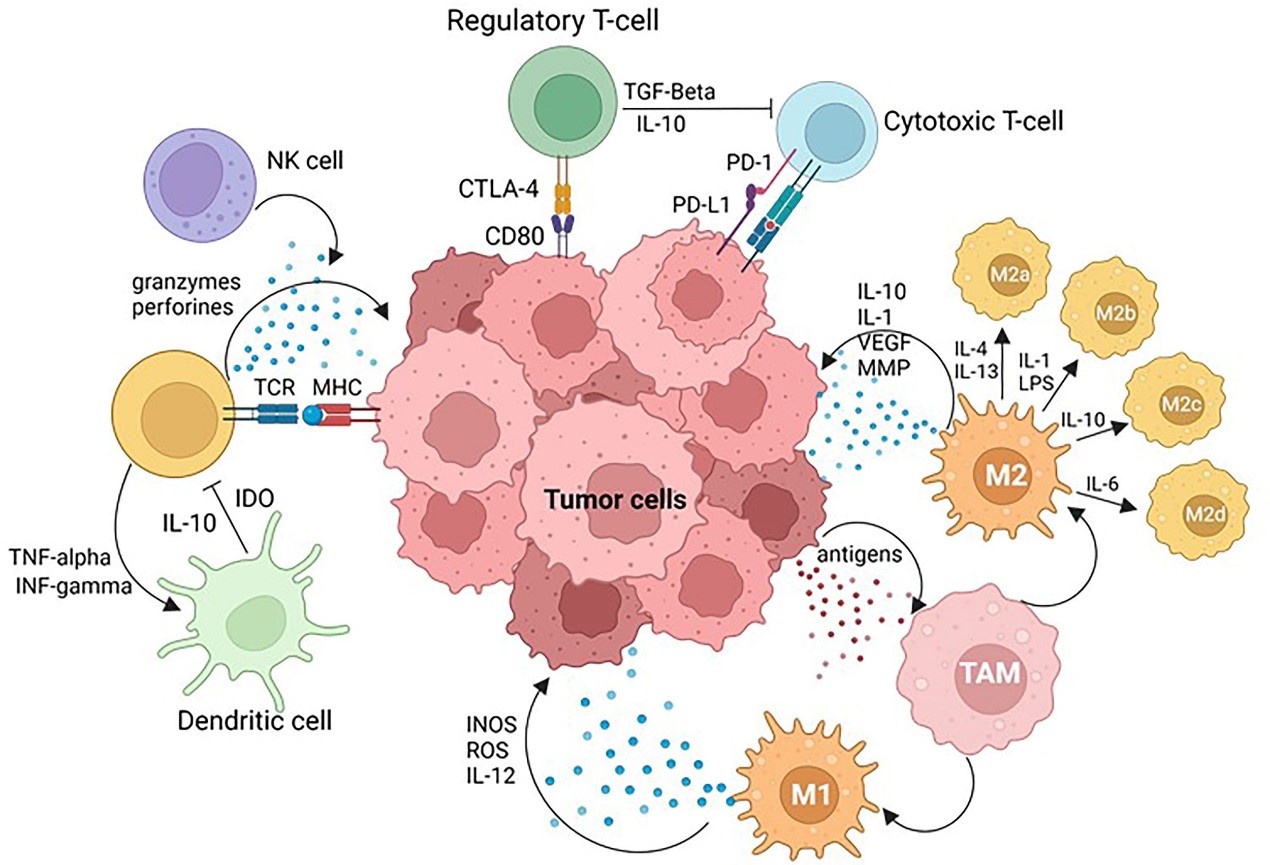
The interaction between cancer and the host immune system mainly occurs in the reactive tissue stroma surrounding tumors, an area known as the Tumor Immune Microenvironment (TIME). The breakthrough clinical application of immune checkpoint inhibitors, particularly targeted therapies against Programmed Death Protein 1 (PD-1, a T-cell co-inhibitory receptor) and Programmed Death Ligand 1 (PD-L1, also known as B7-H1/CD274), has not only propelled cancer immunotherapy into a new era but also made functional analysis of TIME a core focus in both research and clinical settings. Precise characterization of TIME's cellular composition, functional states, and molecular interaction networks is a crucial prerequisite for screening predictive biomarkers for immunotherapy response. Furthermore, deeply clarifying the dual pro-cancer/anti-cancer mechanisms of various immune cells in TIME and their dynamic associations with tumor cells can provide core support for developing novel immunotherapeutic strategies. However, the high heterogeneity of TIME and the challenge of detecting low-abundance immune molecules pose severe challenges to traditional analytical techniques, urgently requiring highly sensitive, multiplex-targeted in situ detection tools to overcome this dilemma.
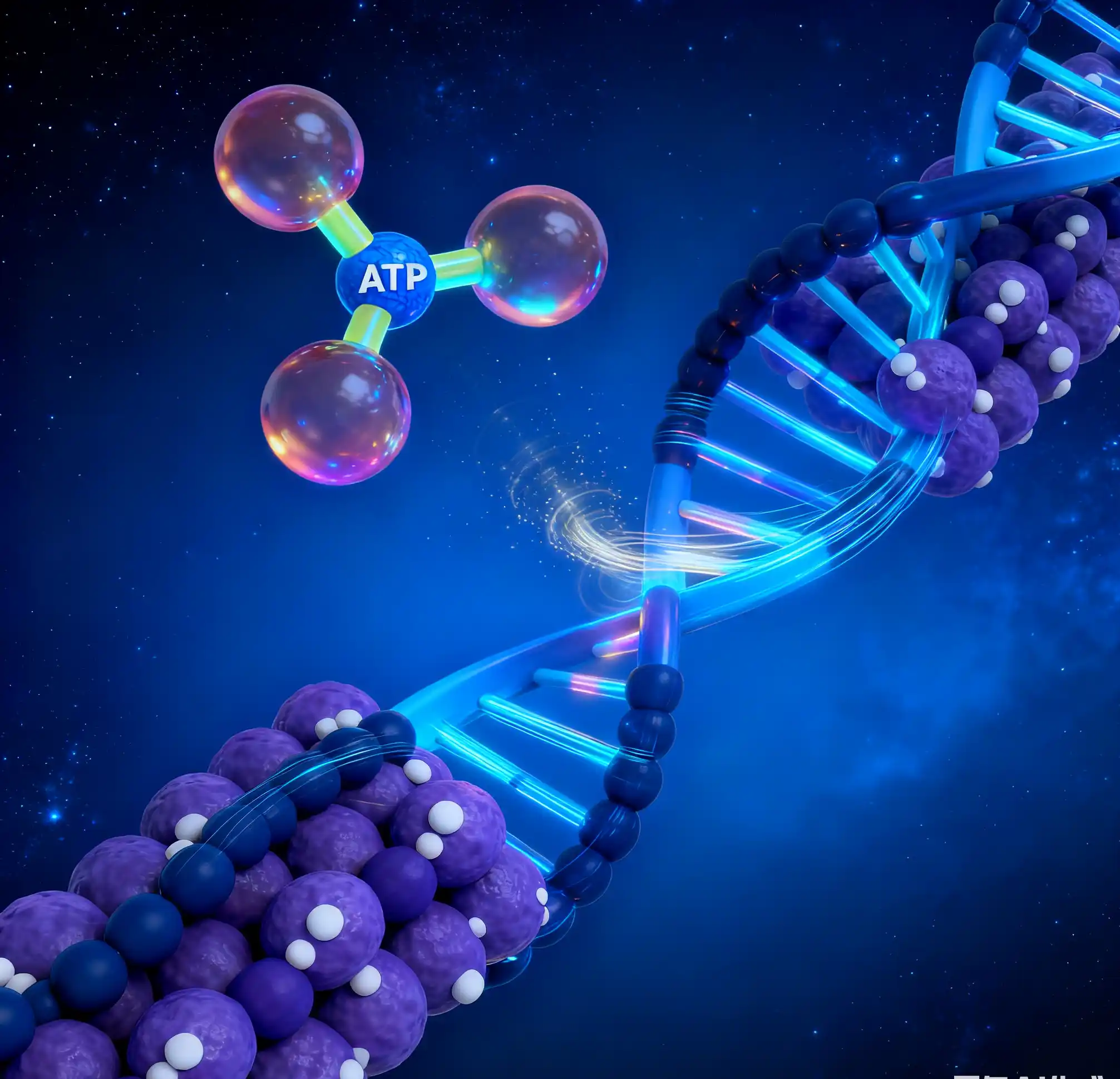
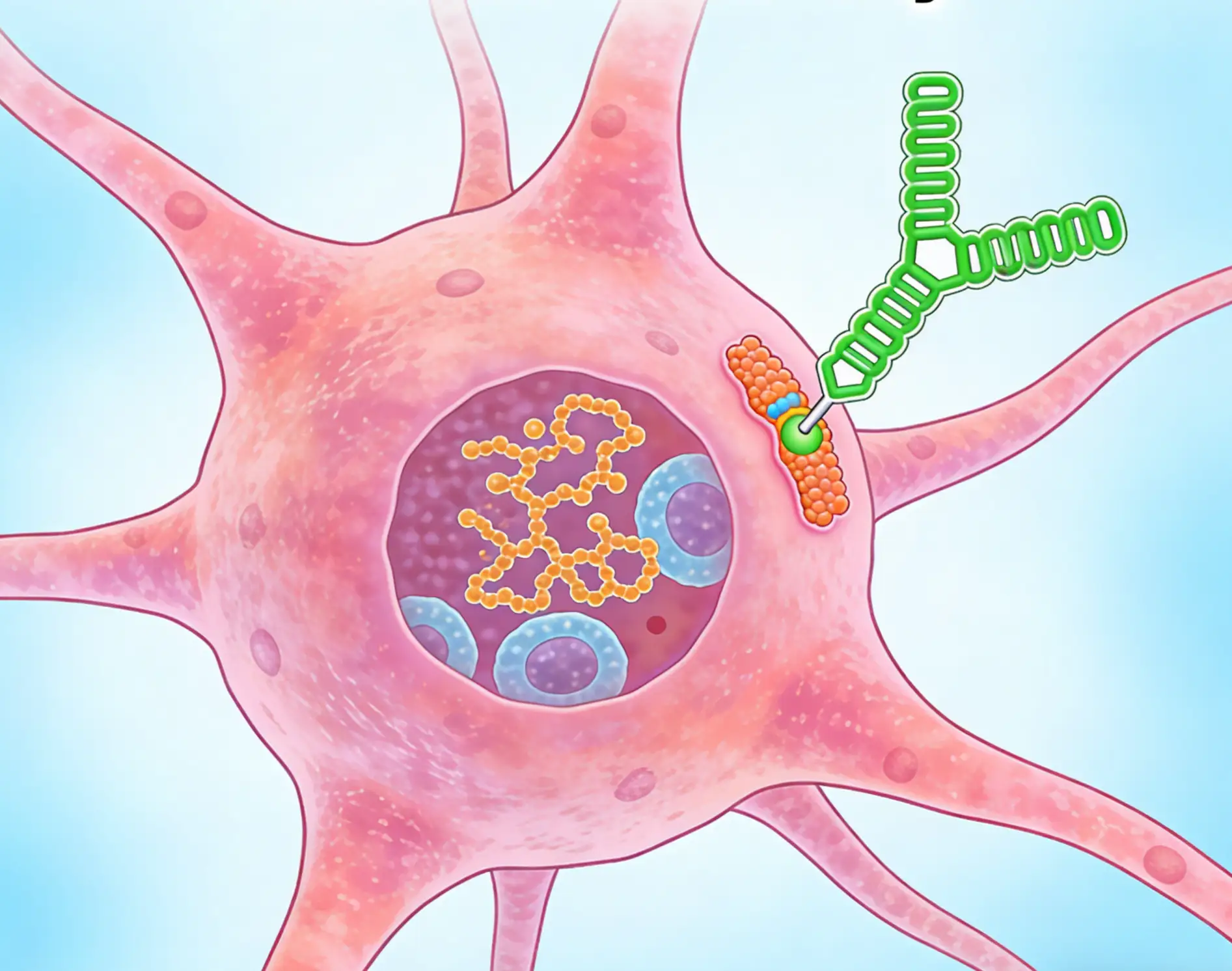
The modulation of chromatin structure is an essential component in the regulation of transcriptional activation and repression. Modifications can be made by at least two evolutionarily conserved strategies, through the disruption of histone-DNA contacts by ATP-dependent chromatin remodelers, or by histone tail modifications including methylation and acetylation.